Animal Imaging
We are pleased to offer digital radiography, ultrasound, thermal imaging, and endoscopy services as a means of providing excellent care to our patients.
Animal Imaging
Animal Imaging
Digital Radiographs
Radiographs (x-ray images) are produced by directing X-rays through the part of the body we are interested in learning more about. The image is produced by how many of the x-rays go through the body depending on its density. Bones and metal are the most dense and leave a white image on the screen whereas soft tissue absorbs varying degrees of x-rays producing shades of gray on the image. Air is black as the x-rays pass right through! Radiographs are a common diagnostic tool used for many purposes including evaluating heart size, looking for abnormal soft tissues, finding fluid in the lungs, assessment of organ size and shape, identifying foreign bodies, and assessing orthopedic disease by looking for bone and joint abnormalities.
Sometimes a mild sedation may need to be given to your animal to help them hold still as movement will make the radiograph blurry!


Digital Ultrasound
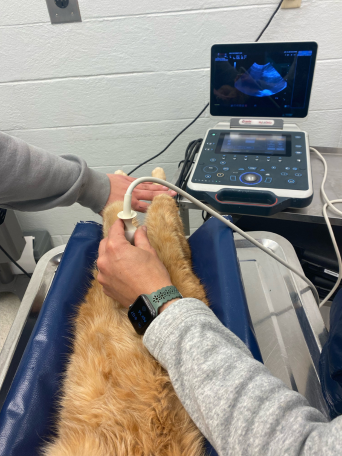
Ultrasound is used to evaluate soft tissues, such as internal organs, and allows us to detect issues in structure and function that cannot be seen with a radiograph. Ultrasound is often combined with radiographs to give us a more complete picture of what is going on inside your pet.
Depending on what type of ultrasound exam is being performed your animal may need to be sedated and have the hair clipped in order to get the best video and pictures possible!
Thermal Imaging
Thermal imaging uses a highly sensitive infrared camera to capture the electromagnetic energy released as varying temperatures from a patient’s body. Veterinary-specific software converts these temperatures into images where the differences in temperatures can be easily visualized by assigning different colors to them. Temperature data directly correlates to changes within the circulatory, nervous, and musculoskeletal systems.
Normal blood flow in animals results in the same surface temperature on each side of the body. Unhealthy patients will have un-equal temperatures from one side of their body to the other. This occurs due to blood flow and neurologic changes in tissue at a diseased location. Unexpected warmer or cooler areas, or asymmetry within the patient, tell us to look more closely at that region for a potential problem!
Increased temperatures (hyperthermia) are a direct result of increased blood flow. This may be due to inflammation, infection, or malignancy. Decreased temperatures (hypothermia) are a result of decreased blood flow. This is usually due to a neurological condition resulting in vasoconstriction.
Sometimes a thermal image can help us identify problems in your pet before you see any sign of them at home! A thermal image is a non-invasive and quick way to get inside information that can help prevent and treat problems. It can easily be added to any wellness or sick animal exam and is a great way to track changes over time by comparing images from one exam to the next!


Endoscopy
Endoscopy involves the use of either a rigid or flexible fiberoptic instrument to allow visual examination of the internal organs without invasive exploratory surgery. A small camera at the end of the endoscope provides images of the internal structures of the body, allowing the veterinarian to view disease processes and take biopsies for further diagnostics. It can often be used for foreign body removal as well. This procedure allows for a non-surgical alternative in some cases for pets that minimizes anesthesia time and allows for a shorter recovery and hospital stay.
